
The greatest notable change between agonists in the structure of the protein results from a shift in the hydrogen bonding capability of Asn293, which, when bound to BI167107, hydrogen bonds the "amide carbonyl on the head group of BI167107" (Ring et al., 2013). The chemical structures of HBI and epinephrine lack this amide group, leading to pressure on the protein to reestablish this bond by bending the Asn293 side chain. This leads to greater shifts in the overall conformation of the protein, including a shift of His296 and a conformational change in the third extracellular loop, leading to a surface change (Ring et al., 2013).
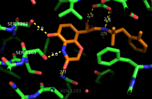
This figure shows the main binding polar contacts of BI167107 (orange)
to the adrenoceptor(green). The catechol-analog head forms specific
contacts with two serine residues and an asparagine residue.
great pyMOL pictures on this page- they are really clear
ReplyDeleteThe pictures with the white background are really good to see and I like the use of colours to highlight the most important residues!
ReplyDeleteI would have put a white background for the text as well because even though I like the background image it makes it a bit hard to read.
Information is clear, but a bit small, which makes it harder to read.
ReplyDelete